

PyMOL结构显示的公开科研资料图片,保留原作者、许可证与来源。示意图、历史资料和实验图片按原图说明识别。
原始图片说明
Proteasome cutaway PyMol.png
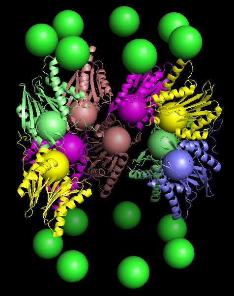
Semi-schematic of the proteasome (PDB accession code 1G0U). The alpha subunits of the two outer rings are shown in green as 5 Angstrom spheres centered on the subunit centroid, which coincides with the CB atom of Leu137. The beta subunits of the two inner rings are shown as ribbon diagrams of different colors. The CA atoms of residue 1 of the beta subunits (corresponding to the catalytic residues) are likewise shown as 5 Angstrom spheres of the same color as their ribbon. The beta subunits of 1G0U corresponding to chain H, M, N, 1 and 2 have been removed to produce a "cutaway" effect; thus, there are five subunits in the lower beta ring and four in the upper. The sphere size was set using the "set sphere_scale=X" command in PyMol. Made by me on 5 January 2007 using PyMol; the cutaway PDB file was made by hand editing. Hereby released under the GFDL.
图片用途与识别
本图用于科研资料浏览和学习,实际设备、样本和操作条件以原始图注及实验记录为准;原理示意、历史器材、书籍封面或软件界面不能当作本网站在售商品实物。使用图像作研究或采购参考时,应同时核对型号、尺度、版本与完整来源。
作者与使用许可
作者:No machine-readable author provided. WillowW assumed (based on copyright claims).
许可:CC BY-SA 3.0