

共聚焦显微成像的公开科研资料图片,保留原作者、许可证与来源。示意图、历史资料和实验图片按原图说明识别。
原始图片说明
Superficial spreading melanoma on dermoscopy, reflectance confocal microscopy and histopathology 2.jpg
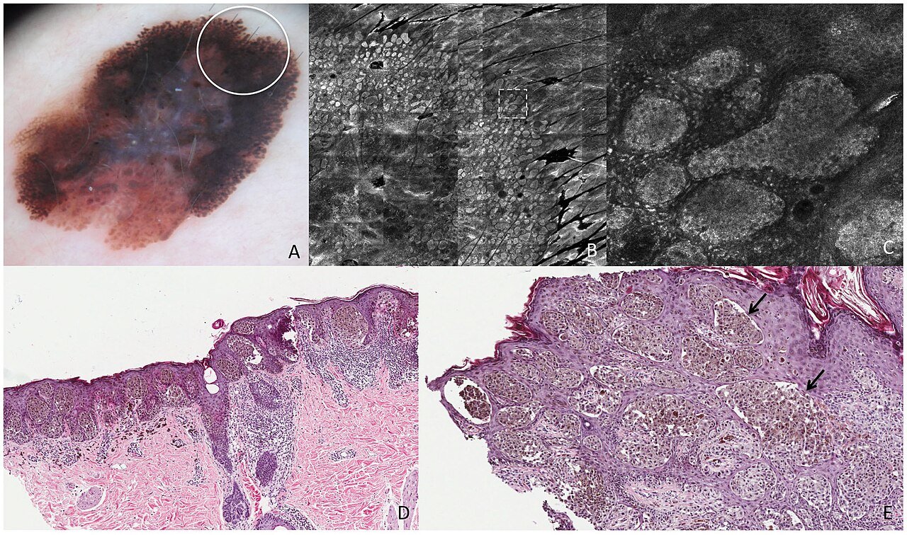
Superficial spreading melanoma on dermoscopy, reflectance confocal microscopy and histopathology: Breslow thickness 0,79 mm. This lesion shows on dermoscopy (A) pseudopods (white circle corresponds to the punch area). RCM mosaic image (B, 363 mm) at the level of the DEJ shows compact aggregates of atypical cells distributed in a linear arrangement toward the periphery with a dense nest at the extremity (the area inside the dashed square is represented in figure C). RCM individual image (C, 0,560,5 mm) at the level of the DEJ shows a pseudopod in detail, characterized by elongated, dense and bright peripheral aggregate. Perpendicular section (D) shows nests of atypical melanocytes distributed contiguously toward the periphery along the DEJ. Transverse section (E) shows nests of atypical cells arranged in a linear manner throughout the periphery of the lesion (black arrows).
图片用途与识别
本图用于科研资料浏览和学习,实际设备、样本和操作条件以原始图注及实验记录为准;原理示意、历史器材、书籍封面或软件界面不能当作本网站在售商品实物。使用图像作研究或采购参考时,应同时核对型号、尺度、版本与完整来源。
作者与使用许可
作者:Juliana Casagrande Tavoloni Braga, Mariana Petaccia Macedo, Clovis Pinto, João Duprat, Maria Dirlei Begnami, Giovanni Pellacani, Gisele Gargantini Rezze
许可:CC BY 4.0